Common Breast Problems
Breast Lump
You or your doctor may find a breast lump by looking at or feeling your breast. It is difficult to determine by examination alone if a lump is caused by breast cancer. Although most breast lumps in women age 20 to 50 are not cancerous, all new breast lumps should be evaluated by a doctor to determine if further testing is needed.
Evaluation
After a breast examination, the best test for evaluating a breast lump depends, in part, upon your age.
Women under age 30
If you are under 30 years and you find a lump before your menstrual period, you may be advised to have a repeat breast examination after your period has ended. In this age group, breast lumps are often caused by hormonal changes and will resolve after your menstrual cycle.
If the lump does not go away when your period is over, you will likely need further testing with a breast ultrasound or needle aspiration biopsy to determine whether the lump is fluid filled or solid. Mammograms are not usually performed in women under 30 years old. MRI will be more informative in this age group. MRI can only be ordered by your breast specialist.
Women age 30 and older
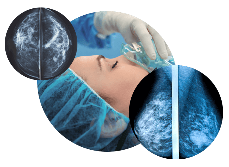
Women who are age 30 or older who find a new breast lump will need a diagnostic mammogram, and usually an ultrasound, as well. During a diagnostic mammogram, a mammography technician works with a radiologist to study the area that feels or appears abnormal. If the lump appears suspicious on the mammogram and/or the ultrasound, a breast biopsy is usually recommended.
We recommend you visit your breast specialist to have a proper breast examination and review of your images, to ensure that no extra biopsy are needed in case of discovering additional lesions.
Breast biopsy
A breast biopsy is usually recommended to further evaluate a new breast lump. A breast ultrasound, mammogram, or MRI may be recommended before a biopsy.
Abnormal mammogram
Having an abnormal screening (initial) mammogram can cause concern. Fortunately, most women with abnormal mammograms do not have breast cancer. An abnormal mammogram may be due to a mass, a collection of calcium deposits (called calcifications), or other factors.
- If you have an abnormal screening mammogram, the next step depends on the type of abnormality found.
- If the abnormality is benign (not cancerous), you may be advised to have a follow up mammogram in six months
- If the mammogram shows an area that is abnormal, the next step is to have additional imaging, with a diagnostic mammogram. During a diagnostic mammogram, a mammography technician works with a radiologist to study the area that feels or appears abnormal. The radiologist can usually review the mammogram immediately and discuss the results with you. In some cases, an ultrasound may be performed to better define the abnormality.
- In many cases, the diagnostic mammogram shows that the abnormality is benign (not cancerous), and no further testing is needed. However, if the diagnostic mammogram is indeterminate or suspicious for cancer, a breast biopsy is recommended.
Breast pain or tenderness
The most common type of breast pain is caused by the hormones that control the menstrual period. These hormonal changes can cause pain in both breasts several days before the menstrual period begins. Because the pain can come and go with the menstrual cycle, it is called “cyclical” breast pain. Cyclical breast pain is not usually caused by breast cancer or other serious breast problems.
Less commonly, a woman can have breast pain that does not come and go with the menstrual cycle (also called noncyclical breast pain). This type of pain is not related to the menstrual cycle and might occur in only one breast or one area of the breast. Noncyclical breast pain is usually caused by a problem outside the breast, such as muscle or connective tissue strain, skin injury, spinal conditions, or problems in another organ system (eg, heart burn, chest pain). Noncyclical breast pain is caused by breast cancer in only a very small percentage of women.
If you are worried about breast pain, speak to your doctor to determine if you need further testing. If testing shows no signs of a serious problem, you can try one or more of the following treatments:
- Pain relief medicines, such as panadol or ibuprofen. Women with very severe breast pain are sometimes treated with a prescription medicine.
- Decrease the dose or stop taking medicines that contain estrogen (after a discussion with your healthcare provider).
- Wear a well-fitted support or sports bra.
- Consider making changes to your diet. Elimination of caffeine and a low fat, high complex carbohydrate diet is helpful for some women. Dietary supplements such as vitamin E and evening primrose oil have also been suggested for breast pain, however, this is not effective in everyone.
Nipple discharge
Having a milky-colored discharge (also called galactorrhea) from both nipples is common, especially during the first year after giving birth. Nipple discharge from both breasts can also occur in women with an underactive thyroid (hypothyroid), as a side effect of certain medications, or because of a growth in the pituitary (a part of the brain), causing an increase in a hormone called prolactin.
As with other ducts in the body, breast ducts make and carry secretions. Many women can express (squeeze out) a small amount of yellowish, greenish, or brownish discharge. This is often called “physiologic” discharge and is not a cause for concern. Physiologic discharge is not bloody.
Spontaneous nipple discharge (discharge that occurs without squeezing) or nipple discharge that is clear or bloody may be caused by an abnormal growth within the breast or, less commonly, by breast cancer.
Any woman with nipple discharge should be evaluated by a healthcare provider. A mammogram, breast ultrasound will be recommended for initial evaluation.
Inverted nipples
Many women are born with nipples that naturally invert (pull in) at times and evert (poke out) at other times. Other women find that this happens after breast feeding. Nipple inversion of this type is not cause for concern.
If your nipples have always been everted, however, and begin to invert for no obvious reason, this should be evaluated by your healthcare provider. Most causes of nipple inversion are not a cause for concern, but occasionally this is the first sign of a breast cancer. New nipple inversion is usually evaluated with a breast examination and mammogram as a first step.
Breast skin changes
Skin problems can develop on or near the breast, some of which cause itching, scaling or crusting, dimpling, swelling, redness, or changes in skin colour. While most of these changes are not caused by a serious breast problem, it is important to be evaluated if a skin problem on your breast does not resolve within a few days.
More serious causes of skin changes on the breast can include less common forms of breast cancer, such as Paget disease or inflammatory breast cancer. Other, more common skin problems, such as rashes, moles, cysts, or skin infections can occur on the skin of the breast, as well.
The evaluation of breast skin changes usually includes a breast examination and may include a mammogram. A skin biopsy may be needed to confirm the diagnosis.
When to seek help
If you find a new breast problem, you should make an appointment with your doctor within a few weeks. Although breast problems are not usually an emergency, delaying the evaluation for months can potentially allow the problem to worsen.
In some cases, this evaluation will be all that is needed. In other cases, you may be referred for further testing or evaluation with a breast surgeon.
If your initial evaluation shows no sign of a problem but you remain concerned, talk to your healthcare provider. Further testing, follow-up over time, or referral to a breast specialist may be recommended.
Where to get more information
Your healthcare provider is the best source of information for questions and concerns related to your medical problem.